
Manchester Analysis
Manchester
Morphological Analysis
&
Shoeing
“The gait abnormality created by a specific lesion is the gait abnormality that will cause the lesion.”
(James R. Rooney, Biomechanics of lameness in horses, 1969)

Thursday, February 4th at 10,30pm: Manchester walked his first steps onto the Science of Motion’s property. The transport from Canada was effectuated by Rick Bodi Transport (514) 591- 6223. Manchester had to be transported in a box-stall due to his size and a history of colic when traveling. The transport company kept us informed all the way as to their progress and Manchester’s health. Their arrival in Florida was right on schedule.
Manchester's arrives in Florida
Manchester walked out of the trailer in good condition. He was tired as expected after such a long trip, but only moderately stressed and in good health. As he arrived closer to the barn Manchester was greeted by one of the resident horses and responded peacefully. It was reassuring to see that Manchester was interested and alert. Even more comforting was the fact that when he walked into his new stall Manchester investigated both feed and water buckets and started to drink immediately. Sometimes after the stress of a long trip, horses stop drinking, putting themselves at risk of dehydration.
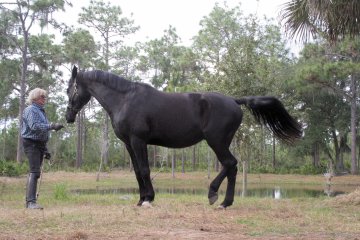
For the next day’s photo session, Manchester posed willingly. We did not ask him to stand square like for an in-hand presentation. We asked for several halts and selected the pictures that illustrated his most natural posture.

He naturally stands with the hind legs relatively far behind him, which is not a comfortable posture for sickle hocks. To allow comparison, we placed him in a posture more appropriated for his type of hind leg conformation.

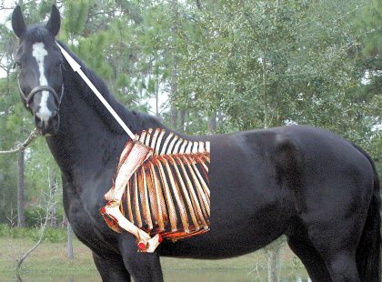
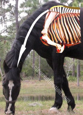
Horses do not voluntarily stress stifle, hocks, or any other limbs’ joints unless they have to protect an even greater strain in another part of their body. Perhaps the site of greater discomfort may be more forward in the spine. Following the line of Manchester’s thoracolumbar column, a dorsal curvature of the spine (roach-back or kyphosis) is apparent in the junction between thoracic and lumbar vertebrae. Under his long coat, the kyphosis is not really obvious. The dorsal curvature is easier to see once the horse has been clipped.

In a survey of 443 cases exhibiting disorders of the thoracolumbar spine, Leo Jeffcott suggested that kyphosis was, in young horses, the result of a protective posture. “Kyphosis was most frequently seen in young horses exhibiting varying degrees of bilateral stifle damage. Improvement only occurred if the underlying cause was resolved.”(1980)
Often the dorsal curvature became less apparent when the surrounding muscle mass increases. The thought that the kyphosis might be related to stifle issues may apply to Manchester since in some instances the swing phase of his left hind leg suggests such pain. However, while it is probable that the roach back may became less apparent when the back muscles become stronger, it is improbable that, at Manchester’s age, the dorsal curvature of the spine could be totally corrected.
As we look at the line of Manchester’s spine ahead of the kyphosis toward the whither, the vertebral column’s orientation is down hill. This is not due to the horse’s conformation, which is in fact up hill. The visual impression is due to the lack of muscular development. Also, the trunk is suspended too low between the forelegs. This is due to insufficient strength of the forelegs’ extrinsic muscles. This lack of muscular development is probably the reason why Manchester stands with the hind legs behind him. The posture is therefore more the result of improper muscular development than specific pain in another area of his body. In fact the pain is likely to be in the fetlock, hocks and stifle joints of the rear legs.
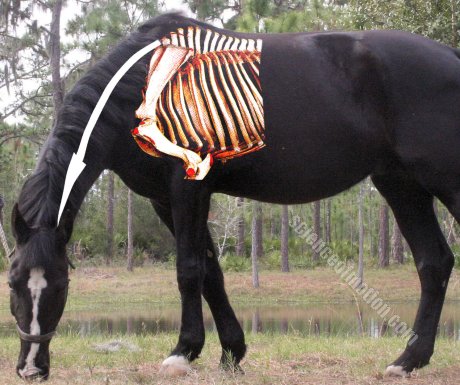
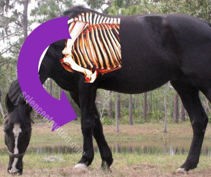
The left picture is the way Manchester looks today. In the right picture we show through computer manipulation how the back muscles will look in several months. As well as how he will stand over the front legs.
Often, the horse’s adaptation to excessive weight on the forelegs is to shift the front limbs backward underneath the body. Similar reaction applies when the hind legs are unable to resist forward movement of the body over the forelegs. Actually, Manchester tends to hold the forelegs backward underneath him.
From the perspective of traditional thinking, it might be surprising to read about the hind legs resisting forward movement of the body. A brief explanation might be necessary. From alighting and during the initial 45% of the stride, the hind leg on support is developing a shock absorbing force, resisting accelerations of gravity and inertia. This sequence of the support phase is referred to as the braking phase. By contrast, the second half of the stride is defined as the pushing phase. The hind legs are then producing a force in the direction of the motion.
The stance of the forelegs presents the same succession of braking and pushing activity. However, two differences occur. One is that the forelegs’ braking phase is shorter than the hind legs. It lasts only 40% of the support phase. The second difference is that the forelegs are producing essentially an upward propulsive force. This explains why recent measurements have demonstrated that the forelegs were producing more upward propulsive activity than the hind legs. “In horses, and most other mammalian quadrupeds, 57% of the vertical impulse is applied through the thoracic limbs, and only 43% through the hind limbs.” (H. W. Merkens, H. C. Schamhardt, G. J. van Osch, A. J. van den Bogert, 1993).
The dynamic of the braking and pushing activities is easier to understand by analyzing a horse at the piaff. The braking activity of the hind limb on support is the sequence where the fetlock, the hock, the stifle and the hip joints are folding. The pushing activity occurs when the joints are extending. The flexion of the joints that characterize the braking activity is used to resist gravity and inertia forces and consequently, forward displacement of the body over the forelegs, which in turn allows the forelegs to develop greater vertical impulse. During piaff, “The hind legs have a considerable braking activity to avoid forward movement of the body over the forelegs The forelimbs have a larger propulsive activity.” (Eric Barrey, Sophie Biau, Locomotion of dressage horses Conference on Equine Sports Medicine and Science - 2002)
Since the pushing force of the hind legs is the sequence of the stride where the joints extend, one can understand that during piaff, the horse reduces the propulsive activity of the hind legs to the minimum. An energetic hind legs’ propulsive action would generate elevation of the croup and consequently, forward shift of the body’s weight over the forelegs.
Training techniques that attempt to stimulate greater propulsive activity of the hind legs by acting on the lower legs with a dressage whip are counterproductive since they are stimulating the wrong reflexes. Activating the hind legs, the dressage whip stimulates greater propulsive activity of the hind legs and consequent elevation of the croup. Such elevation shifts the body weight forward over the forelegs, hampering the front limbs’ ability to propel the horse’s body upward.
“Creative minds have always been known to survive any kind of bad training.” (Anna Freud) The horses’ creativity is demonstrated on this video. We compare a horse trained to the piaff in respect of actual knowledge of the equine physiology, with the performances executed during the Atlanta Dressage Olympics by horses, which for a large percentage, are trained with the dressage whip activating the hind legs. A large majority of the horses are dealing with the croup elevation shifting the forelegs backward underneath themselves and consequently became unable to sustain rhythmic elevation of the forelegs.

During the photo session, Manchester became interested in a patch of grass in front of him. He lowered and elongated the neck demonstrating a great ability to shift the forelegs backward underneath him.
When a horse lowers the neck, the traction exerted by the nuchal ligament on the tip of the dorsal spinous processes of the thoracic vertebrae induces a verticalization of the dorsal spinous processes. “In the living horse, as is also the case in anatomical specimens, the lowering of the neck produces a verticalization of the spinous processes in the wither.” (Jean Marie Denoix, Spinal biomechanics and functional anatomy, 1999)
The term verticalization does have a French accent but is descriptive. In the wither area, the inclination of the dorsal spinous processes is oriented backward. As the neck lowers, the traction exerted by the nuchal ligament on the tip of dorsal spinous in the wither induces a rotation of the vertebrae, thereby placing the dorsal spinous processes in a more vertical position.
If the muscles that support the trunk between the forelegs are strong enough to resist sagging of the trunk, the rotation of the cranial thoracic vertebrae has two positive effects. One is to induce flexion of the thoracic spine. The other is to separate the dorsal spinous processes. The separation of the processes has been measured and found to be greatest between the 6th and 10th thoracic vertebrae. “Intervertebral movements were greater between T6 and T10, where the average amount of flexion was 1.7º” (Denoix)
In a different study, the greater range of movement has been found to be between T2 and T8. (K. S. Gellman. E. J. A. Bertram, The equine nuchal ligament: structural and material properties.)
Such separation of the dorsal spinous processes is possible in the wither area because the ligament which connects the tip of the processes remains somewhat elastic until about the 10th thoracic vertebra. The ligament in question is named the supraspinous ligament. It is in fact the continuation of the nuchal ligament which connects the skull to the 4th thoracic vertebra. As the ligament continues all along the tip of the thoracolumbar’s neural spines (dorsal spinous processes), it becomes the supraspinous ligament. The nuchal ligament is elastic (elastic fibers), while the supraspinous ligament is not (collagen fibers). However, the conversion from a large percentage of elastic fibers, to a majority of collagen fibers is progressive. Some elasticity remains in the wither area allowing the tip of the neural spines to move apart.
The nuchal ligament in illustrated here in white. The gradual loss of elasticity of the supraspinous ligament is illustrated here in yellow and brown.
The separation of the dorsal spinous processes that occurs in the wither area when the horse’s neck is lowered, can be used as a therapy when the horse is subject to impingement of the dorsal spinous process (kissing spine) in this specific area.
The verticalization of the dorsal spinous processes in the wither area also has several negative effects, one of which is increased weight on the forelegs. When the muscles supporting the trunk from the front legs do not have sufficient strength, the rotation of the vertebrae created by the traction of the nuchal ligament on the tip of the dorsal spinous processes increases the load on the forelegs.
Backward shift of the forelegs underneath the horse’s body does not occur solely during piaff. We have observed in many instances that such backward shift was initiated by the lowering of the neck and consequent verticalization of the dorsal spinous processes. These observations stand as our working hypothesis since, as of yet, no specific studies has been made on the matter.
One of the horses which initiated these observations was a young Hanoverian suffering from a condition known as contracted tendon. As it is often the case, the problem commenced when the horse was 6 months old. The farrier worked on the hoof to avoid the development of club foot. The horse shoer also used different techniques aiming at stimulating elongation of the tendons. While obtaining great successes on the development of the hoof capsule, the technique did not have the expected results on the tendons. The horse cancelled the effects of the corrective shoeing by lowering the neck and rotating the cranial thoracic vertebrae. Doing so, the horse kept the weight over the coffin bone, avoiding tension on the flexor tendons.
Lowering of the neck and the consequent rotation of the vertebrae in the wither engendered in the young horse, stiffening the back muscles in the caudal thoracic and lumbar region. Such muscular stiffening was treated with massage therapy. Also, the rotation engendered a vertical rotation of the scapula which was treated through chiropractic manipulation. The horse counteracted the effects of all therapies by rotating even further the cranial thoracic vertebrae.
At age three, the horse’s owner observed that the forelegs were gradually shifting backward. As a last resort surgery was executed, cutting the check ligament of the right front leg. The surgery provided improvement but did not resolve the lameness issue.
Through all these corrective therapies and manipulations the horse associated human contact with physical pain. His revolt exploded with the backing process. The horse arrived at the training center with the reputation of being difficult. The next day, as I was planning to walk him quietly around the farm, the horse demonstrated that the term “difficult” was an understatement. The horse’s reaction illustrates both his mental and physical state. Once forward movement was regained, the lameness of the right hind leg was apparent. It took a few months to recreate physical comfort and the horse turned into a very well balance athlete, both, physically and mentally.
CLICK HERE FOR VIDEO OF CASE OF CONTRACTED TENDON
The horse was extremely heavy on the fore hand and the balance issue needed to be addressed before any efficient gymnastic could be done. As soon as the young horse realized that the gymnastic program was creating physical comfort instead of pain, his willingness and ability to process mentally was quite good. Balance control was created, educating the biomechanical properties of the vertebral column and placing the neck into a long but higher posture.
The horse was very intelligent and in a surprisingly short period of time, the young horse realized that proper use of his vertebral column mechanism was more efficient and more comfortable than rotating the cranial thoracic vertebrae and shifting the weight on his front toes. Proper balance restored soundness and the horse no longer stood with the front legs shifted backward under his body.
The stance of Manchester’s front legs that we have created through the magic of computer manipulation is one of our goals. In a few months we truly expect to show a non edited picture of Manchester standing with the front legs at the right place.

The view from behind shows a pelvis turned to the left as well as rotated transversally.
The left side and the left hip are higher than the right side and the right hip.
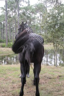
From the hock down, both hocks are oriented toward the medial line.
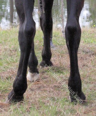
There is a noticeable inflammation of both fetlocks and both pasterns are oriented toward the outside.
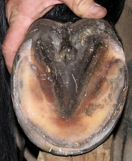
As expected, the hoof capsule was deformed with the medial half of the hoof much smaller and under-run toward the frog.
For long, the veterinary world has regarded back issues as the consequence of upper or lower legs problems. A more advanced school of thought is now considering possible interactions between upper and lower legs issues and back muscle imbalance. “Limbs disorders are often treated exclusively, without investigating possible structural and functional interactions between the spine, upper limb and lower limb.” (Kevin K. Haussler, DVM, DC, PhD) preface of the Veterinary clinics of north America, Equine Practice, Vol 15, Number 1, April 1999) The Science of Motion resolutely regards the biomechanical properties of the horse’s vertebral column as the source of all body’s movements and consequently most limbs’ kinematic abnormalities and subsequent injuries. However, no efficient work can be made recreating symmetry of the back muscles or other issues if the horse does not have a sound platform, that is, if the hooves are not properly balanced.
One perspective could be that Manchester’s hock morphology caused the deviation of the pasterns toward the outside and heavy impact on the inside wall. The direction of the forces that such abnormal impact induces on the upper legs and vertebral column could explain the torsion of the thoracolumbar spine and consequent deviation of the pelvis. Such hypothesis would suggest that appropriated shoeing may have prevented the development of the problem. At theoretical level such hypothesis is possible. Another thought could be that the torsion of the thoracolumbar column and consequent lateral and transversal displacement of the pelvis may have engendered the lower limbs’ abnormalities. Once again, and from a theoretical point of view, the thought is possible. However, the truth is probably a combination of both.
The hocks may have been morphologically too close and consequently the pasterns and the growth of the hooves may have taken the direction observed on this primary analysis. The abnormalities of the lower legs may have engendered biokinematic abnormalities of the thoracolumbar spine and pelvis, which in turn have aggravated the stresses on the lower legs. Horses do not work through problems by thinking about long term effects. Instead, they focus on instant relief. In Manchester’s case, as is very often the situation, the compensations that the horse developed to an initial abnormality created greater problems over a period of time. James Rooney’s statement is now easy to understand. “The gait abnormality created by a specific lesion is the gait abnormality that will cause the lesion.”
Whatever perspective one is inclined to believe, no proper gymnastic of the back can be achieved without a sound platform, (shoeing). As well, no adjustment of the lower legs can succeed without addressing the kinematics as well as dynamic abnormalities of the horse’s vertebral column that induced excessive or abnormal stresses on the lower legs. Manchester does need a team work approach.
The phenomenon is true for every aspects of the equine industry. A perfectly appropriated therapy will be effective only if the horse’s physical education addresses the problem that necessitated the therapy in the first place. Hoping that shock wave or other therapy will fix the problem and riding the horse the same way once lameness has vanished will simply lead the horse to the same type of injury.
We asked Mike Galagher if he would be willing to join the team. Mike has extensive shoeing experience. For many years, Mike’s skill allowed hunter jumpers to enter the ring sound after having exited the previous ring lame or questionable. Later Mike realized that an even better application of his skill, knowledge and experience would be to fully restore soundness. Mike observed Manchester in motion, as well as standing still. Two options were possible: a gradual adjustment or a drastic change. Considering that no real gymnastic would be efficient without a sound support of the horse’s lower legs, we decided in favor of an aggressive approach. Such decision necessitated that we give Manchester the time he needs to adjust to his new base.
Manchester’s Shoeing
By Mike Gallagher
“I observed Manchester’s stance and noticed that the right hock was close to the median and that the hoof was splayed out to a severe degree, possibly as much as 40º. I then observed the left hind leg and saw basically the same thing. The animal also displayed very deformed hoof capsules, under-run toward the frog from the medial, and running out from the frog toward the lateral, on both.
Watching the horse walk several steps from behind, it became apparent that each leg displayed a different affectation to the abnormal. It became clear that a balancing platform needed to be applied. To immediately help relieve his discomfort and allow his recovery program to ensue, a radical approach was called for. We decided on a shoe that in essence disregarded the hoof capsule and positioned the shoe where the hoof should have been normally. The steel shoe when affixed protruded from the inside wall a half inch or more.

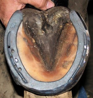
This rather unsettling aspect of the appliance had almost an instant stress relieving effect, up the inside of the hind legs from the floor to the top line of the rump. This was made apparent by Manchester quickly indicating his preference to bare all his weight on the first [right] leg shod. The left leg performed the same way after its shoeing.
Left picture is before shoeing. Right picture is after shoeing.
The lateral side of the shoes were beveled at a 45 degree angle and fit very close [tight] to try to eliminate the debilitating leverage that had developed there over a period of time. Both shoes installed, Manchester further displayed a more normal stance, with legs more apart and an apparent leveling through the point of the hips.
To our relief when Manchester was walked he showed a marked change to the normal stride use of both legs. The shoes should allow easier break- over. Also the length of the branch past the heel will support the bony column far more than an unshod foot.”
(Mike has since completed his report going in greater details about the shoeing technique applied. Anyone interested in Mike’s views can contact him by phone, Mike Galagher: (863)438 0801
Mike spent literally a full day with Manchester. He worked with the horse all afternoon, stayed overnight to allow Manchester to rest, and finished the job the next morning. When it was time for the invoice Mike told us that his time, knowledge, material and skill were his donation to Manchester’s project.
I rode Manchester shortly after his shoeing. I stayed at the walk and was able to appreciate the difference. I had clearly in my mind the memory of the feeling that Manchester gave me when I rode him a few months earlier in Quebec. It was at this time a collapse in the caudal thoracic and lumbar region on the right side. My impression at that time was that while the left hind may have been the original issue, the compensation of the right side was the main problem. In the airplane flying back home I even theorized a gymnastic program.
The feeling is now completely different. The extension of the right lumbar region no longer exists. In fact, Manchester is holding the lumbar region a little too round but equally on both sides. A glitch can be felt on the left side, at the beginning of the left hind leg’s swing phase.
Manchester was cautious about his movements and as planned, we gave him the time to adjust to his new platform, walking him on long reins. Each day, Manchester’s back felt a little different. After four days at the walk, the glitch on the left side was no longer perceptible.
Manchester and I need more time together, walking, trotting and soon cantering in the country side before I can decide about the reeducation program and where to start. This will be the subject of our next discussion.
Jean Luc Cornille ©2010
www.scienceofmotion.com
jeanluc@scienceofmotion.com
